
The concept of balance, defined by Webster as "a state of equilibrium," permeates various aspects of life, from financial budgets to personal well-being. In the realm of equine athletics and general horse welfare, this principle of equilibrium is not merely philosophical but fundamentally critical, especially concerning joint health. Maintaining the intricate balance within an equine athlete’s musculoskeletal system is paramount for their longevity, performance, and overall quality of life. Without this equilibrium, the carefully constructed "equine team"—comprising the horse, owner, trainer, and veterinarian—faces significant challenges, with the horse’s well-being always at the core.
The Equine Team: A Foundation of Balance
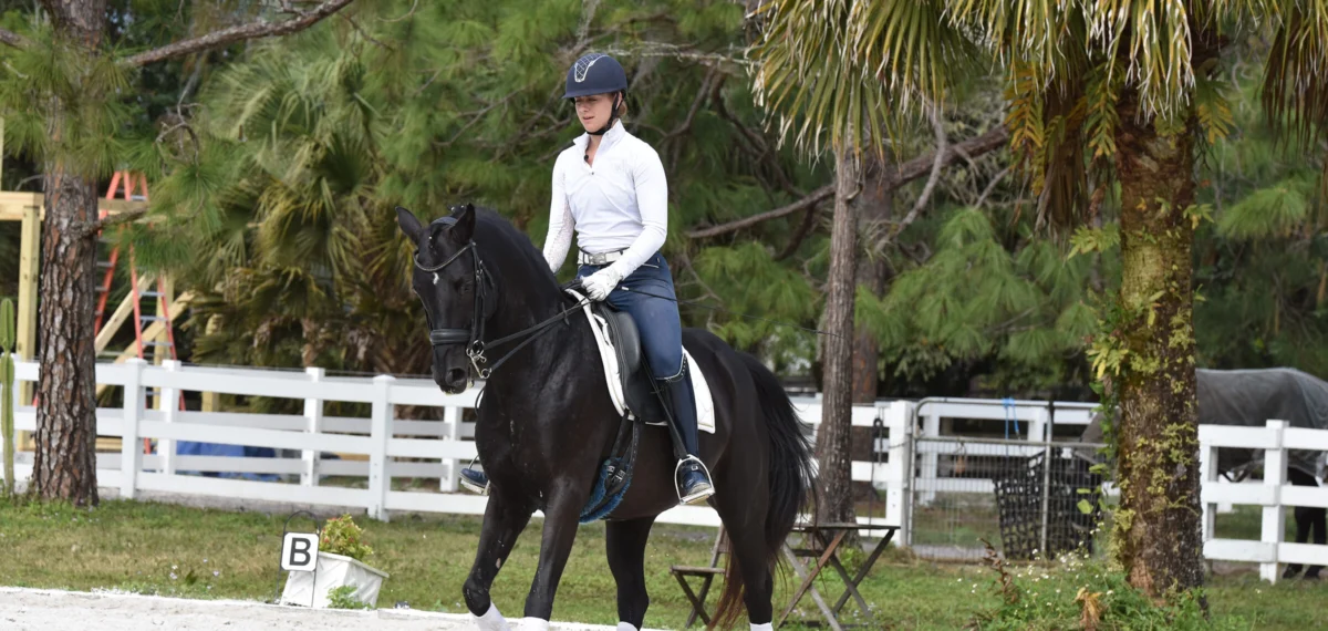
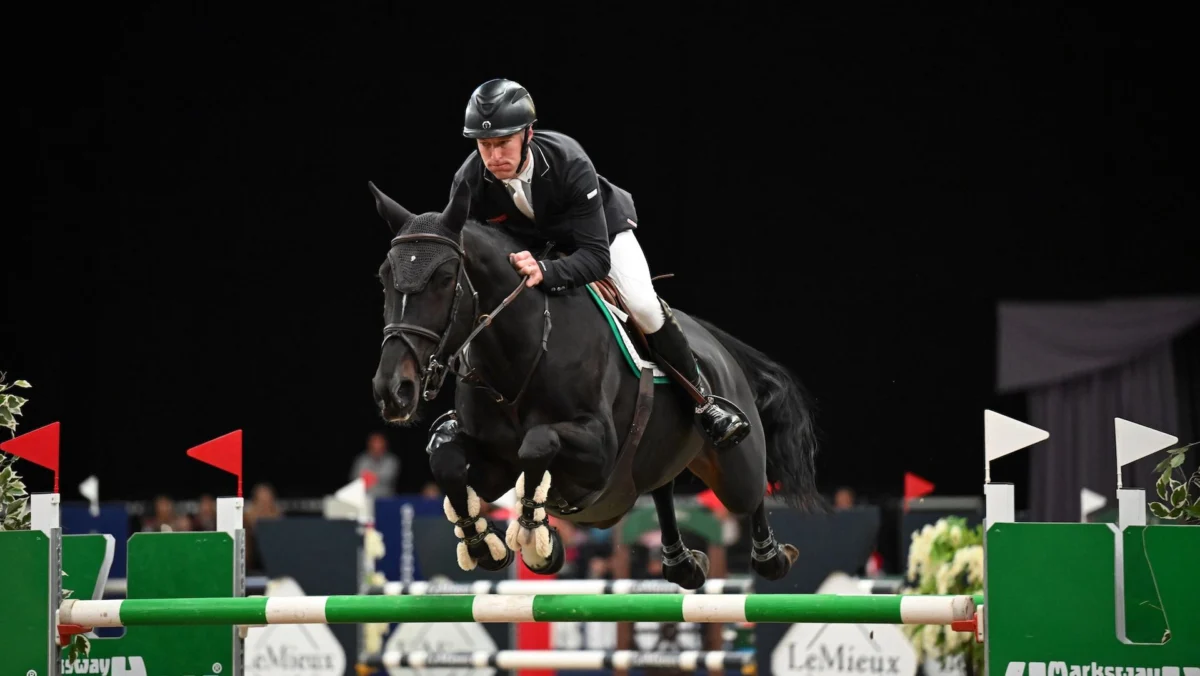
At the heart of any successful equine endeavor is the horse itself. Prioritizing the horse’s welfare and making decisions that are unequivocally in its best interest forms the bedrock of a balanced and effective team. This commitment ensures sustained success and prevents short-sighted choices from compromising long-term health. Following the horse, the owner plays a pivotal role. Without the owner’s dedication, resources, and commitment to responsible care, the entire system falters. The trainer guides the horse’s physical development and performance, while the veterinarian provides essential medical expertise, diagnostics, and treatment. When these four elements function in a cohesive, balanced manner, the horse stands the best chance of remaining sound, healthy, and successful. Conversely, a disruption in any part of this equation can precipitate a cascade of negative consequences, often manifesting as lameness.
Equine lameness is a pervasive issue within the equestrian world. Studies consistently indicate that lameness accounts for a significant proportion, often exceeding 60%, of all health problems in performance horses, leading to substantial economic impact through veterinary costs, lost competition time, and diminished market value. For instance, a report from the American Association of Equine Practitioners (AAEP) highlighted that musculoskeletal issues, predominantly lameness, are the leading cause of economic loss in the equine industry, underscoring the urgency of effective joint health management.
Understanding the Complex Equine Joint
Far from being a simple "vat of lubricating fluid" akin to a car engine, the equine joint is a marvel of biological engineering. It is a highly complex, dynamic structure designed to facilitate movement, absorb shock, and withstand immense forces. Key components include:

- Subchondral Bone: The bone directly beneath the articular cartilage, providing structural support and playing a crucial role in load bearing. It adapts to stress, becoming thicker and stiffer with increased loads, but excessive, unrecovered stress can lead to microfractures and pain.
- Articular Cartilage: A smooth, resilient tissue covering the ends of bones within the joint. Its primary functions are to reduce friction during movement and distribute mechanical loads evenly across the joint surface. Healthy cartilage allows for virtually frictionless motion.
- Synovial Fluid: A viscous, lubricating fluid contained within the joint capsule. It nourishes the articular cartilage (which lacks its own blood supply) and provides further shock absorption and lubrication. Its quality, particularly its hyaluronate concentration, is critical for joint health.
- Joint Capsule: A fibrous sac enclosing the joint, containing the synovial fluid.
- Peri-articular Soft Tissues: This category includes tendons, ligaments, and muscles surrounding the joint. Ligaments provide crucial stability, preventing excessive or abnormal movement, while tendons connect muscle to bone, transmitting force. Muscles surrounding larger joints like the shoulder and hip contribute significantly to stability.
Any disease or damage to even one of these components can profoundly affect overall joint function. For example, a strained collateral ligament will destabilize a joint, potentially leading to secondary cartilage damage, while inflammation of the synovial membrane (synovitis) can alter synovial fluid composition, reducing its protective qualities.
The Imperative of Accurate Diagnostics
Given the intricate nature of equine joints and the myriad causes of lameness, precise diagnosis is not just beneficial but absolutely essential. Relying on tradition, fear, or anecdotal evidence for treatment decisions can not only be ineffective but also detrimental, leading to prolonged suffering for the horse and significant financial strain for the owner. A comprehensive diagnostic approach typically involves:
-
Initial Lameness Examination: This fundamental step involves observing the horse at rest and in motion (walk, trot, canter) on various surfaces (hard, soft, straight lines, circles).
- Flexion Tests: Specific joints are held in flexion for a short period before the horse is trotted off. An exacerbation of lameness often indicates pain within the flexed joint or its associated structures.
- Palpation: Careful manual examination of the limbs and joints to detect heat, swelling, pain response, or altered conformation.
- Hoof Testers: Used to localize pain within the hoof, which can often be a primary or secondary cause of lameness.
-
Diagnostic Anesthesia (Nerve and Joint Blocks): Once preliminary observations localize the lameness to a general region, local anesthetic agents are injected around specific nerves or directly into joints. If the lameness resolves after a block, it precisely pinpoints the source of pain.
-
Advanced Imaging Modalities:
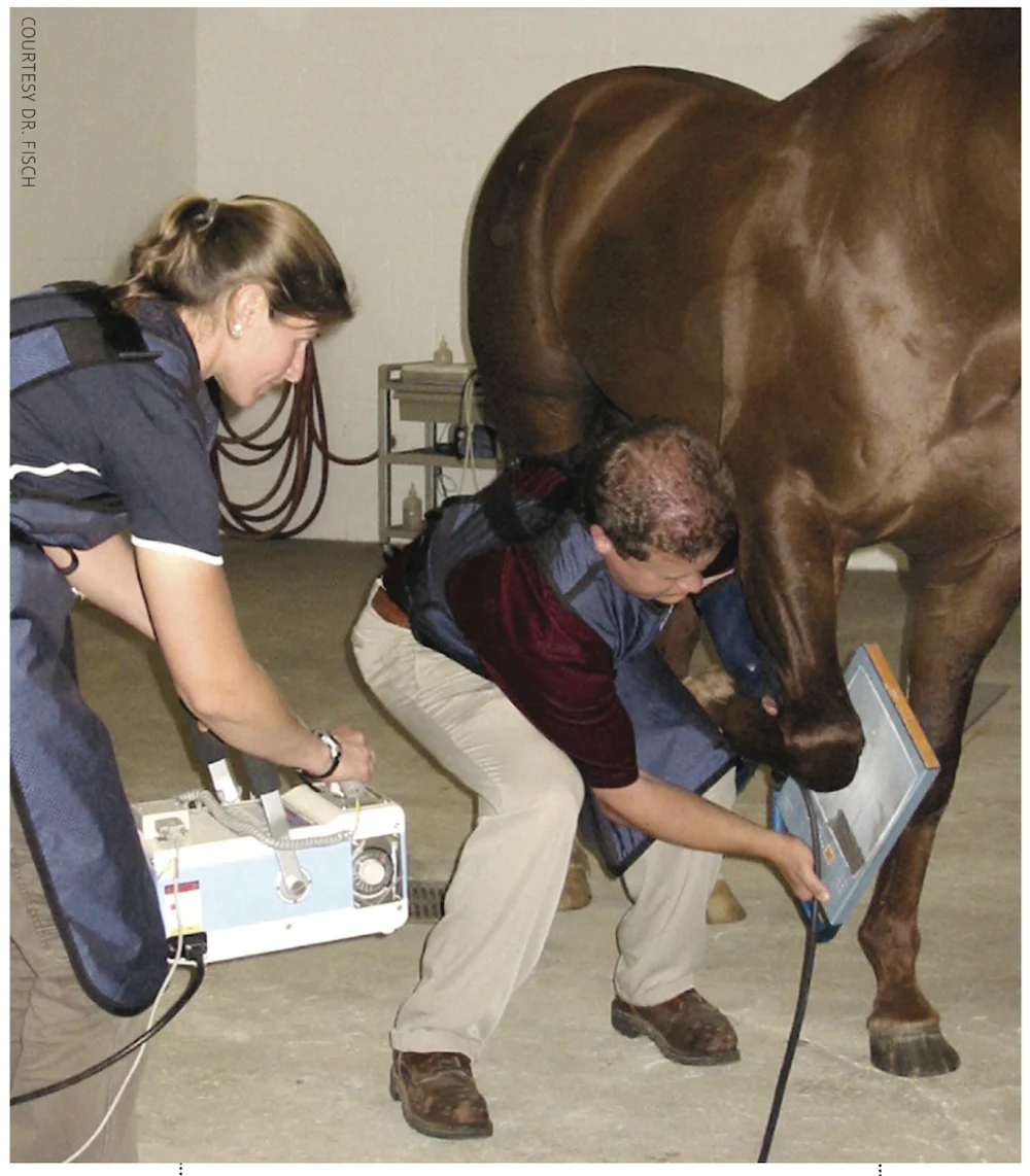
- Radiography (X-rays): High-quality digital radiographs are invaluable for assessing bone changes, such as osteoarthritis, fractures, bone cysts, or subchondral bone disease. While excellent, they may not always reveal soft tissue or early cartilage changes.
- Ultrasound: Primarily used to visualize soft tissue structures like tendons, ligaments, joint capsules, and menisci (in stifle joints). It’s crucial for diagnosing strains, tears, and inflammatory changes.
- Magnetic Resonance Imaging (MRI): Considered the gold standard for diagnosing complex lameness, especially in the foot and lower limb. MRI provides detailed images of both bone and soft tissue, revealing subtle lesions not visible on X-rays or ultrasound.
- Computed Tomography (CT): Offers highly detailed, three-dimensional images of bone structures, useful for complex fractures or specific bone conditions, particularly in the head, neck, and lower limbs.
- Scintigraphy (Bone Scan): Utilizes a radioactive tracer that accumulates in areas of increased bone turnover or inflammation, highlighting "hot spots" that indicate active pathology, even before radiographic changes are evident.
As Dr. Stephen Fisch, Dr. Chad Baumwart, and Dr. Patrick First emphasize, thorough evaluation extends beyond the immediate site of lameness to the horse’s overall body condition, diet, and training regimen. A holistic view is critical because problems in one area inevitably impact others over time.

Injecting Balance: When and How to Treat
Joint injections represent an important therapeutic tool in equine veterinary medicine, but their judicious application is paramount. When used at the correct time, with an accurate diagnosis, and with appropriate medications, injections can significantly improve joint health, alleviate pain, and extend an equine athlete’s career. However, misused or ill-timed injections can cause more harm than good.
Types of Joint Injections and Their Nuances:
- Corticosteroids: These potent anti-inflammatory agents are widely used to reduce pain and inflammation within joints. However, the choice of steroid is critical. Chondroprotective steroids (e.g., triamcinolone acetonide, betamethasone) are generally preferred as they have been shown to have minimal or even beneficial effects on cartilage. Conversely, some less soluble crystalline steroids (e.g., methylprednisolone acetate) can persist in the joint and may be associated with long-term cartilage degradation, especially in high-motion joints. The debate surrounding steroid selection and frequency is ongoing, with veterinarians continually balancing immediate pain relief with long-term joint health.
- Hyaluronic Acid (HA): A natural component of synovial fluid, HA acts as a lubricant and anti-inflammatory agent, improving synovial fluid quality and cartilage health. It can be injected alone or in combination with corticosteroids.
- Biologic Therapies: These cutting-edge treatments harness the horse’s own healing mechanisms:
- IRAP (Interleukin-1 Receptor Antagonist Protein): Targets and blocks the inflammatory cytokine interleukin-1, which is highly destructive to cartilage in osteoarthritis.
- PRP (Platelet-Rich Plasma): Concentrated platelets from the horse’s own blood contain growth factors that promote tissue healing and reduce inflammation.
- Stem Cell Therapy: Involves injecting mesenchymal stem cells (often derived from fat or bone marrow) into damaged joints, aiming to regenerate tissue and modulate inflammation. While promising, research into their efficacy for specific joint conditions is ongoing.
The decision to inject should always be part of a comprehensive treatment plan, not a standalone solution. Risks, though relatively low, include joint infection (a severe complication requiring immediate and aggressive treatment), flare reactions, and the potential for masking underlying issues if the diagnosis is incomplete.
Beyond Injections: A Spectrum of Therapies
Joint injections are one component of a broader management strategy. Other crucial treatments and considerations include:
- Rest and Controlled Exercise: For many soft tissue injuries (ligament strains, muscle tears, bone bruising), rest is the most critical therapy. This may range from stall rest to controlled hand walking or small paddock turnout, followed by a gradual return to work. Impatience in this phase is a common cause of re-injury.
- Systemic Medications: Non-steroidal anti-inflammatory drugs (NSAIDs) like phenylbutazone or flunixin meglumine are often used orally or intravenously to manage pain and inflammation, particularly in acute phases. Oral joint supplements (e.g., glucosamine, chondroitin sulfate, MSM) are widely used, though scientific evidence for their efficacy varies.
- Physical Rehabilitation: A growing field in equine medicine, rehabilitation programs utilize various modalities such as therapeutic laser, shockwave therapy, hydrotherapy (underwater treadmill), massage, chiropractic adjustments, and acupuncture to improve strength, flexibility, proprioception, and reduce pain.
- Farriery and Hoof Balance: Proper hoof care and shoeing are foundational to biomechanical balance. Imbalances in the hoof can significantly impact joint loading and contribute to lameness throughout the limb. A skilled farrier working in conjunction with the veterinarian and trainer is invaluable.
Training, Stress, and Recovery: The Preventative Pillars

The adage "prevention is better than cure" holds immense truth in equine joint health. A carefully planned training regimen that incorporates appropriate stress and sufficient recovery is vital to preventing repetitive microtrauma, a leading cause of osteoarthritis.
- Beneficial Stress vs. Overload: Bones and cartilage adapt and strengthen in response to controlled, progressive loading, adhering to principles like Wolff’s Law (bone remodels in response to the stresses placed upon it). This adaptive process makes tissues more resilient. However, excessive loading, particularly at high frequency without adequate recovery, exceeds the tissue’s capacity for repair and leads to micro-damage accumulation.
- Recovery Time: This is a frequently overlooked but critical component of any training program. For racehorses, for example, a minimum of 10-14 days is often required for bones and joints to fully recover after a high-speed workout or race. Pushing horses too hard, too often, without this recovery window, inevitably leads to cumulative damage and lameness. The same principle applies to performance and pleasure horses in disciplines involving repetitive movements or high impact. Trainers must meticulously balance intensity and volume of work with adequate rest and recovery periods, incorporating varied training, cross-training, and active recovery strategies.
- Arena Footing and Surfaces: The type and condition of training surfaces significantly impact joint loading. Hard, uneven surfaces increase concussion, while excessively deep or inconsistent footing can place undue stress on soft tissues. Maintaining optimal arena footing is a key preventative measure.
Holistic Management: Diet and Lifestyle
The horse’s overall health, diet, and environment profoundly influence joint well-being.
- Nutrition: A balanced diet providing essential vitamins, minerals, and high-quality protein is fundamental. Specific nutrients, such as omega-3 fatty acids (found in flaxseed or fish oil), can have anti-inflammatory properties that support joint health. Antioxidants help combat oxidative stress within the joint. Careful attention to body weight is also critical, as obesity places excessive strain on joints.
- Supplementation: While many joint supplements are marketed, their efficacy varies. Owners should consult with their veterinarian to ensure any supplements are appropriate, evidence-based, and do not create an imbalance by providing "too much of a good thing."
- Early Recognition of Signs: Owners and trainers are often the first to notice subtle changes in a horse’s gait, attitude, or performance. Early signs of joint disease can include decreased range of motion, stiffness, reluctance to perform certain movements, or a general dullness. While articular cartilage lacks nerves, pain arises when surrounding structures become inflamed. Therefore, any change warrants prompt veterinary investigation. Decreased viscosity of synovial fluid, detectable through simple tests like the "string test" (where normal synovial fluid strings between fingers), can also be an early indicator.
Broader Implications: Welfare, Ethics, and Industry Standards
The pursuit of balance in equine joint health extends beyond individual horses to broader ethical and economic considerations within the equestrian industry. Prioritizing horse welfare means making decisions that ensure a horse’s long, comfortable, and productive life, rather than pushing for short-term gains at the expense of their health. The economic burden of lameness on owners and the industry is immense, highlighting that investing in preventative care, accurate diagnostics, and balanced treatment plans ultimately leads to cost savings and sustainable participation in equestrian sports.
Globally, leading equine veterinary organizations, such as the AAEP and the British Equine Veterinary Association (BEVA), consistently advocate for evidence-based practices, emphasizing comprehensive diagnostics before treatment and the responsible use of medications. This commitment to higher standards of care benefits not only the horses but also strengthens the public’s trust in equestrian sports.
The Future of Equine Joint Health

Research in equine joint health continues to evolve rapidly, bringing forth new diagnostic tools and therapeutic options. Advances in regenerative medicine, such as more sophisticated stem cell applications and gene therapies, hold promise for repairing damaged tissues and preventing disease progression. Improved imaging techniques offer even greater precision in diagnosing subtle pathologies. The ongoing collaboration between veterinarians, researchers, and industry stakeholders will continue to refine our understanding and management of equine joint health.
Ultimately, the goal is to foster an environment where every member of the equine team – horse, owner, trainer, and veterinarian – operates with a shared understanding and commitment to maintaining equilibrium. This collaborative, fact-based approach, centered on the horse’s best interests, ensures an overall healthier equine population, more successful athletes, and a vibrant, responsible equestrian community for generations to come.
About Dr. Fisch and Colleagues
Stephen Fisch, DVM, Chad Baumwart, DVM, DACVS, and Patrick First, DVM, are dedicated equine practitioners operating a full-service equine hospital and reproductive center in Tallahassee, Florida. Their practice, AVS Equine Hospital, specializes in performance and racehorse lameness, internal medicine, reproductive diagnostics and treatments, and state-of-the-art surgical procedures for their clients and referring veterinarians. More information about their services can be found at avsequinehospital.com.
This article was originally published in the March 2018 issue of Barrel Horse News.